Spinal cord ultrasound implant shows promise in early tests
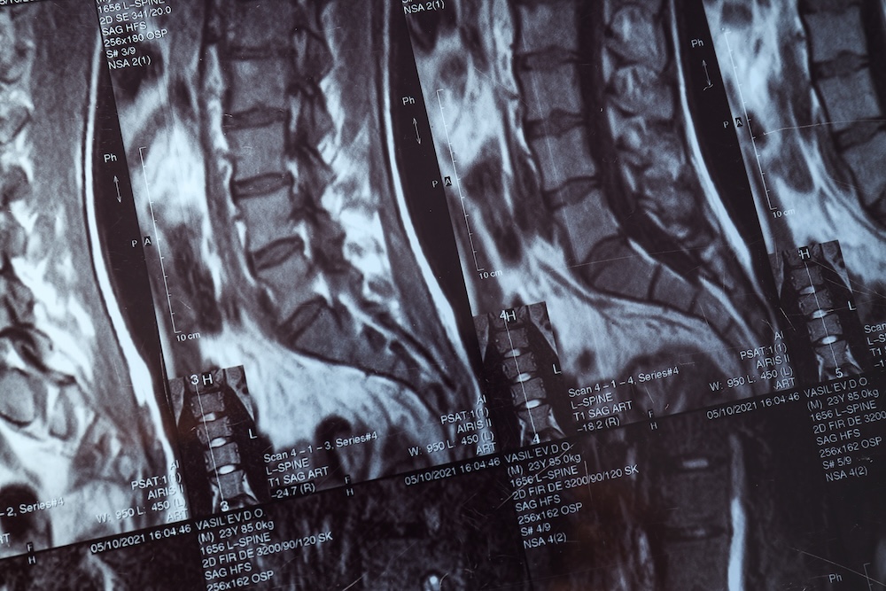
An ultrasound implant the size of a small plaster could help doctors track changes at the site of a spinal cord injury after surgery, early animal research suggests.
The temporary device was successfully implanted and provided real-time images and blood flow information around the injured area.
The findings provide early evidence of its potential for monitoring spinal cord injuries, although further research is needed before clinical use.

Researchers at Johns Hopkins Medicine developed the Multimodal Ultrasound System for Injury Care (MUSIC) following a US$13.48m award from the US Department of Defense.
The device is designed to sit over the spinal cord at the injury site during surgery, allowing clinicians to monitor patients during recovery in intensive care.
Amir Manbachi, associate professor of neurosurgery and biomedical engineering at Johns Hopkins University School of Medicine and principal investigator of the project, said the aim was to track changes directly at the injury site.
He said: “When the biology of spinal cord injury is local, our devices should also be able to track and treat local physiology.”
The implant, approximately the size of a small plaster, combines diagnostic ultrasound imaging with an experimental therapeutic function.
Its imaging component is designed to monitor blood flow and changes in surrounding tissues, including spinal fluid and excess fluid accumulation.
Researchers also hope it could help clinicians assess responses to medication and detect changes beyond the immediate injury site.
This information could guide treatment decisions in the days and weeks after surgery, while reducing the need to move patients for imaging during early recovery.
Rather than relying on scans taken at individual points in time, clinicians could obtain information when needed.
The therapeutic component remains exploratory. Researchers are investigating whether it could identify and respond to changes in blood flow through small vessels around the injury.
The aim is to develop technology that could eventually help reduce complications following spinal cord injury, including stroke, loss of vision and further deterioration in movement.
However, the findings come from an initial animal feasibility study. They do not establish whether the device is safe or effective in people or whether it improves recovery outcomes.
MUSIC received FDA Breakthrough Device Designation in 2023, a status intended to support the development and regulatory review of certain medical technologies.
Nicholas Theodore, a former Johns Hopkins neurosurgeon and current adjunct professor of medicine who developed the device with Manbachi, said the timing of treatment was important in spinal cord injury recovery.
Theodore said the researchers envisaged technology that could help clinicians adjust care after surgery, potentially influencing recovery months or years later.
The study builds on earlier work at the Johns Hopkins HEPIUS Innovation Lab, which investigates electrical, ultrasound and physiological interventions for spinal cord injuries.
Further preclinical research will examine how the device can be improved to monitor blood flow, detect changes in spinal tissues and assess other activity near the injury site.
The team intends the technology to be suitable for people of different ages and with injuries sustained through military service, vehicle accidents, falls or sport.
An estimated 18,000 spinal cord injuries occur in the US each year, with vehicle accidents the most common cause.
The research has received support from the Department of Defense’s Defense Advanced Research Projects Agency (DARPA), the National Institutes of Health and other federal and state funding programmes.
The researchers have also received philanthropic and institutional support.
Manbachi and Theodore are named inventors on patents related to the technology, which are assigned to Johns Hopkins University. Both have disclosed professional or financial relationships with medical technology companies.
Further animal studies will assess the device’s monitoring capabilities and potential role in guiding care before any clinical application can be established.
 Air ambulance blood test detects brain bleeds within minutes