Blood test could help identify brain injuries missed by CT scans
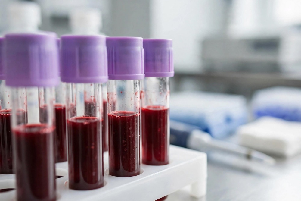
A blood test could help identify brain injuries missed by CT scans, potentially allowing clinicians to recognise patients who need further assessment, new research has found.
Researchers examined blood levels of two proteins to determine whether they could distinguish patients with mild traumatic brain injury (TBI) whose scans show abnormalities from those with no visible evidence of injury.
The findings suggest that one protein, glial fibrillary acidic protein (GFAP), could help identify acute brain injuries within 24 hours, including injuries not visible on CT scans but detected by MRI.

The multi-institutional study, led by researchers at the University of Pittsburgh School of Medicine, investigated whether blood biomarkers could improve the identification and assessment of patients with TBI.
Researchers established clinical thresholds for two proteins released into the bloodstream following brain injury: GFAP and ubiquitin C-terminal hydrolase-L1 (UCH-L1).
The proteins are associated with different types of brain cells and can provide biological evidence of injury.
GFAP is released from glial cells in the brain, while UCH-L1 is released from neurons.
Levels of both proteins rise in the bloodstream immediately after injury before declining over time.
The researchers found that GFAP levels measured within 24 hours could help distinguish patients with acute brain injuries from those with no evidence of abnormalities on brain imaging.
GFAP levels rose sharply between four and 20 hours after injury before gradually declining over the following days.
UCH-L1, by comparison, was less effective at distinguishing between the patient groups examined.
The research draws on TRACK-TBI, a multicentre study collecting clinical information and biological samples from patients attending emergency departments with traumatic brain injuries.
The University of Pittsburgh’s Neurotrauma Clinical Trials Center serves as the study’s primary biorepository, collecting and storing biological samples for analysis.
Previous TRACK-TBI findings showed that more than 50 per cent of participants with mild brain injuries experienced functional limitations one year after their injury.
Shawn Eagle, assistant professor in the university’s Department of Neurological Surgery and lead author of the study, suspects that difficulties identifying injuries contribute to poor long-term outcomes.
He said: “There are amazingly poor long-term outcome rates for people with a TBI who get seen in the emergency department.”
CT scans are generally the first assessment used for patients with suspected traumatic brain injury.
They are widely available, relatively inexpensive and can quickly identify acute injuries, including bleeding inside the skull.
However, CT scans do not detect all forms of brain damage. Approximately 25 per cent of patients with TBI have abnormalities inside the skull that the scans miss.
MRI scans provide more detailed images of brain tissue and can identify injuries not visible on CT, but they are more expensive, take longer and are unsuitable for rapid assessment in emergency departments.
Eagle said: “We’re sending people home with a negative CT scan, but one in four have an abnormality visible on MRI indicating that they should have specialised follow-up care.”
The researchers examined whether blood biomarkers could distinguish patients with positive and negative CT results, including those whose CT scans appeared normal but whose MRI scans showed evidence of injury.
Existing blood tests measuring GFAP and UCH-L1 can already help clinicians determine whether patients with mild TBI require a CT scan.
Results can be available in approximately 15 minutes, potentially avoiding unnecessary imaging.
The latest study investigated whether the same biomarkers could also help identify patients with brain injuries despite a negative CT result.
Eagle said: “We were curious about how the test would perform if you flipped it on its head.”
A point-of-care version of the blood test is now available, allowing whole-blood samples to be analysed at the bedside rather than sent to a laboratory.
This could make biomarker testing more practical in emergency departments.
Eagle said: “We’re working to incorporate this test into the daily clinical practice in the ED.”
The team is also investigating whether blood biomarkers could help predict longer-term outcomes after traumatic brain injury.
The findings suggest that GFAP testing could provide additional information when CT imaging does not reveal an injury, potentially helping clinicians identify patients who may need further assessment and specialist follow-up.
However, the study does not establish that blood testing can replace brain imaging or independently determine which patients require further care.