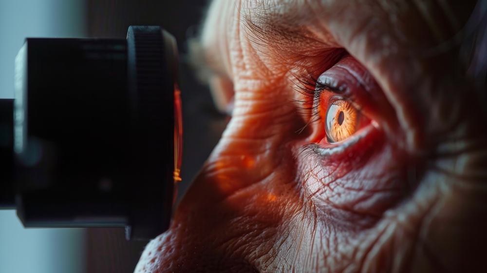
Retina photos could help predict common Alzheimer’s risk factors, new research suggests.
The study found that simple photographs of the retina, the light-sensitive layer at the back of the eye, may offer clues about future brain health.
Researchers said the images can predict many common risk factors associated with developing Alzheimer’s disease.
“We know that Alzheimer’s disease develops over decades, but most of the diagnostic tools focus on late stage pathology when it is too late to intervene,” said Ruogu Fang, professor of biomedical engineering at the University of Florida, who led the study.
“By looking at novel biomarkers, like retinal health, we offer new opportunities to identify patients at risk, offer appropriate tests and encourage them to develop healthy lifestyles to mitigate their risk.”
Biomarkers are measurable signs in the body that may point to disease risk or changes in health.
Fang’s collaborators included Adam Woods, of the University of Florida, and Meta researcher Yunchao Yang. The work was supported in part by the National Science Foundation.
Many patients already have eye photographs taken during routine care, including people with diabetes, glaucoma or cataracts, as well as during some eye tests for prescription glasses.
That makes retinal imaging simpler and cheaper than technologies such as MRI scans, which use magnets and radio waves to create detailed images of the body.
The research team used machine learning, a form of artificial intelligence that finds patterns in data, to assess retinal photographs from more than 40,000 patients in a UK-based patient databank.
They identified areas of the retina linked to Alzheimer’s risk factors, including the arteries and the optic nerve, which carries visual information from the eye to the brain.
“With the assistance of AI, we are now able to identify subtle retinal variations that were formerly overlooked across thousands of subjects, which may function as reliable indicators of future disease risk,” said Seowung Leem, a doctoral student at the University of Florida and first author of the study.
The AI model predicted biological characteristics, including sex and blood pressure, as well as lifestyle factors linked to Alzheimer’s risk, including smoking, alcohol use and insomnia.
Researchers said medical records are often incomplete, while details such as alcohol use and smoking can rely on self-reporting.
Retinal photographs may offer another, more objective way to detect these risk factors.
The images may also capture damage that has built up over years, which can vary between patients who share similar risk factors.
“Retinal morphology could provide measurable indicators of neurovascular integrity, which is highly relevant to Alzheimer’s disease vulnerability,” said Fang, who is also affiliated with the McKnight Brain Institute.
“In this sense, retinal imaging functions less as a surrogate questionnaire and more as an integrated biological sensor of cumulative risk.”
Retinal morphology means the shape and structure of the retina, while neurovascular integrity refers to the health of the links between nerves and blood vessels.
Fang’s group has previously established that retinal photographs can detect active cases of Alzheimer’s disease.
The latest findings focus on whether eye images can help identify risk factors earlier, before irreversible brain damage has taken place.
Researchers said earlier detection could help identify patients who may respond to earlier interventions, including protective lifestyle changes, some medications or brain training.