Imaging technique could let scientists watch Huntington’s disease unfold in real time
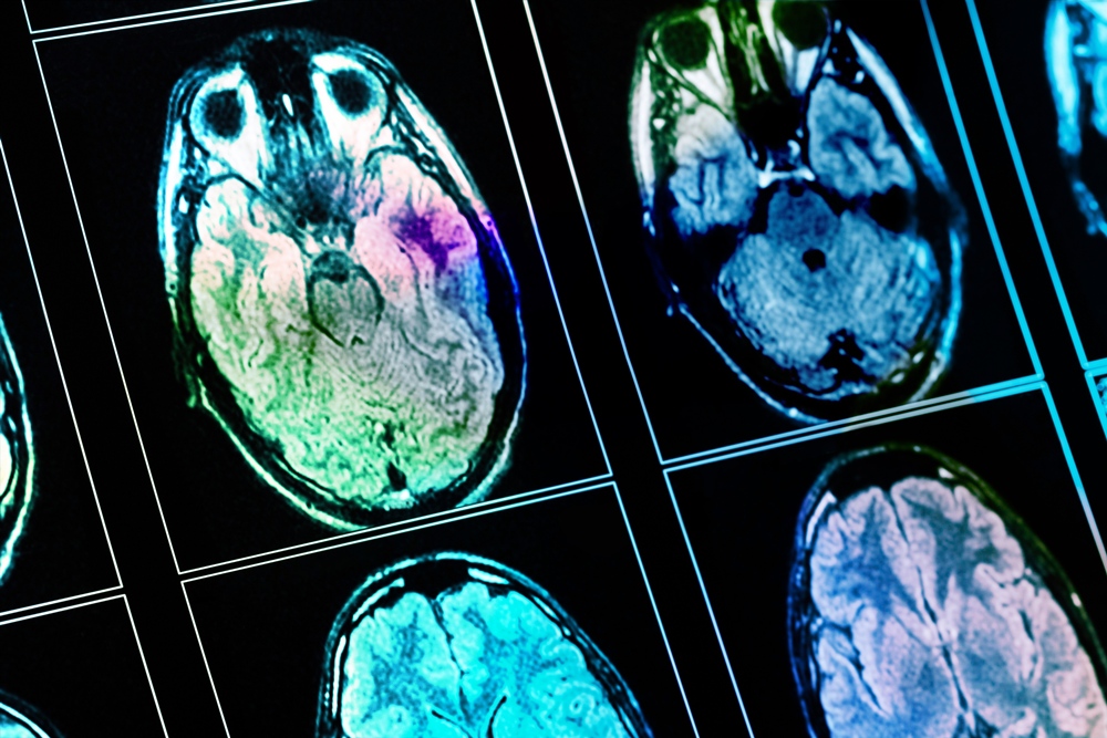
A new imaging technique could help track Huntington’s disease by detecting microscopic changes in brain tissue that conventional scans may miss.
Researchers found the approach could identify abnormalities in brain regions affected by the inherited neurological condition.
The findings could eventually help monitor disease progression and assess experimental treatments, although larger and longer-term studies are needed.

Researchers used an imaging model called Soma and Neurite Density Imaging, or SANDI, to examine brain tissue changes linked to Huntington’s disease.
Huntington’s is an inherited neurodegenerative condition characterised by a progressive loss of cognitive and motor functions.
The disease causes deterioration, or atrophy, in the basal ganglia, a group of brain structures involved in controlling movement, habits and learning.
Conventional MRI scans can measure the volume of brain structures, but cannot directly show the underlying tissue changes that lead to atrophy within the striatum, the signal-processing hub of the basal ganglia.
SANDI is a form of diffusion MRI that analyses how water molecules move inside cells to estimate their size and density.
Researchers wanted to find out whether these measurements could detect Huntington’s-related changes in the basal ganglia and thalami and whether they were linked with movement performance and brain volume.
“We wanted to test how SANDI performs in characterising Huntington’s disease-related abnormalities in the basal ganglia and thalami, and examine associations between SANDI indices, volumetric measurements, and motor performance,” said senior author Claudia Metzler-Baddeley, reader in cognitive neuroscience at the Cardiff University Brain Research Imaging Centre.
“We chose the thalami as control regions based on how neurodegeneration progresses in Huntington’s disease, that is, beginning with early loss of medium spiny neurons in the striatum before extending to the thalamus and other neighbouring structures.
“This is the first time, to our knowledge, that SANDI has been applied in Huntington’s disease, an approach with potential translatability to other neurodegenerative diseases.”
The research was led by Vasileios Ioakeimidis, who was a postdoctoral researcher at the Cardiff University Brain Research Imaging Centre at the time and is now based at the Danish Research Centre for Magnetic Resonance at Amager and Hvidovre Hospital in Copenhagen, Denmark.
The team analysed diffusion MRI data from 56 people with Huntington’s disease and 57 healthy volunteers, all scanned using the same MRI system at the Cardiff University Brain Research Imaging Centre.
Participants with Huntington’s also completed movement assessments, including quick and paced finger-tapping tasks.
Researchers then analysed links between the SANDI measurements, movement performance and conventional measurements of brain volume.
The technique identified differences between the groups in estimates relating to the soma, the rounded body of a cell.
People with Huntington’s had lower estimated soma density, along with higher estimates of soma size and the space between cells, compared with healthy participants.
Researchers said the pattern matched the nerve cell loss and glial reaction previously seen in brain tissue examined after death.
Along with age, the SANDI measurements explained up to 63 per cent of shrinkage within the striatum in Huntington’s disease.
The measurements were also linked to poorer performance in the movement tasks.
As researchers expected, similar changes were not detected in the thalamus.
The researchers said SANDI could provide useful information alongside conventional measurements of brain volume and may be suitable for measuring the effects of disease-modifying treatments in clinical trials.
There are currently no disease-modifying therapies for Huntington’s disease, although several clinical trials are in progress.
Sensitive imaging biomarkers could help researchers assess the effects of new treatments.
However, further research is needed to establish whether SANDI can track disease progression more sensitively than existing imaging biomarkers.
The method would also need to be implemented across the standard MRI systems used in hospitals before it could have wider clinical applications.
“SANDI shows significant promise for tracking Huntington’s disease and testing whether new therapies protect brain cells, and could also be applied to more common neurodegenerative conditions such as Alzheimer’s and Parkinson’s disease,” Metzler-Baddeley concluded.
“Before that can happen, our findings need to be confirmed in larger, long-term studies, and we hope our work will provide a useful framework for those studies.”