


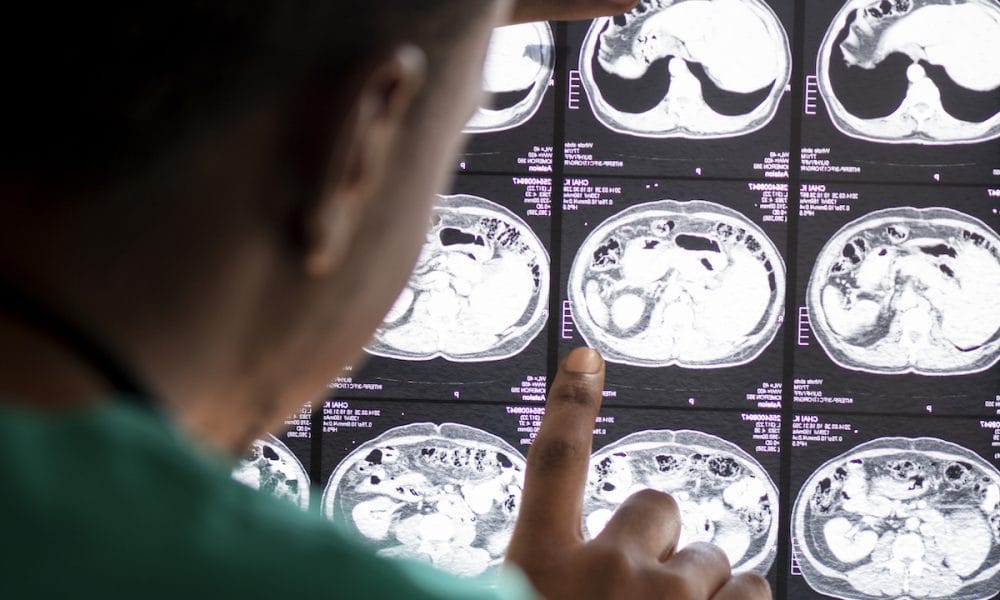
A new MRI technique could improve diagnosis and treatment of multiple sclerosis (MS).
MRI already plays a key role in discovering MS, with the importance being on diagnosing as soon as possible in order to delay its progression.
In the search for ever better methods, a new MRI technique has been used at MedUni Vienna as part of a research project that could pave the way to quicker assessment of disease activity in MS.
Although conventional MRI can detect brain lesions, scientists are researching methods to detect the changes at an earlier microscopic or biochemical stage.
The method, known as proton MR spectroscopy, has been identified as a promising tool for this purpose.
Using this technique, the research group led by Eva Niess (formerly Heckova) and Wolfgang Bogner from MedUni Vienna’s Department of Biomedical Imaging and Image-guided Therapy, working with scientists from MedUni Vienna’s Department of Neurology, went one step further.
The team used MR spectroscopy with a 7-tesla magnet to compare the neurochemical changes in the brains of 65 MS patients with those of 20 healthy controls.

This particularly powerful imaging tool was co-developed by MedUni Vienna researchers and has been used for scientific studies, including of the brain, at MedUni Vienna’s Center of Excellence for High-Field MR since it was commissioned in 2008.
Using 7-tesla MRI, MedUni Vienna researchers have now been able to identify MS-relevant neurochemicals.
“This allowed us to visualise brain changes in regions that appear normal on conventional MRI scans,” says study leader Wolfgang Bogner, pointing to one of the study’s main findings.
According to the study’s lead author, Eva Niess, these findings could play a significant role in the care of MS patients in the future.
”Some neurochemical changes that we’ve been able to visualize with the new technique occur early in the course of the disease and might not only correlate with disability but also predict further disease progression” she said.
More research is needed before these findings can be incorporated into clinical applications, the authors said, adding that the results already show 7-tesla spectroscopic MR imaging to be a valuable new tool in the diagnosis of multiple sclerosis and in the treatment of MS patients.
The method is currently only available on the only 7-Tesla MRI scanner in Austria at MedUni Vienna and only for research purposes, but with significant potential for wider use.
“If the results are confirmed in further studies, this new neuroimaging technique could become a standard imaging tool for initial diagnosis and for monitoring disease activity and treatment in MS patients,” says Wolfgang Bogner, looking to the future.


