Using a single-cell sequencing technique, researchers have created the first cell atlas of focal cortical dysplasia (FCD) – a malformation of the cerebral cortex that leads to drug-resistant epilepsy – paving the way for the development of new specific treatments for this type of dysplasia.
Cases of FCD are most common in children and adolescents, accounting for up to 50 per cent of epilepsy surgeries in this age group.
The atlas has enabled the characterisation of the different cell types present in the brain lesion, and determine which ones are involved in the disease.
Seizures in FCD
The molecular mechanisms responsible for epileptic seizures in patients with FCD are poorly understood. People with severe cases can have between 40 and 50 seizures a day, with loss of consciousness and falls. When medications fail to control the seizures, surgery is an option, but it carries risks, such as vision, hearing and speech problems.
In this study, the researchers were able to map at cellular resolution both transcriptional changes – involved in the process of converting DNA into messenger RNA (the “recipe” for making proteins) – and epigenetic changes (modifying gene expression through biochemical processes without changing the DNA sequence).
These mechanisms regulate how genes are activated or deactivated to produce proteins and other functional molecules, known as gene expression.
The study also identified subpopulations of neurons, microglia and astrocytes involved in the disease. This group of cells forms the cerebral cortex and ensures the functioning, protection and adaptation of the nervous system.
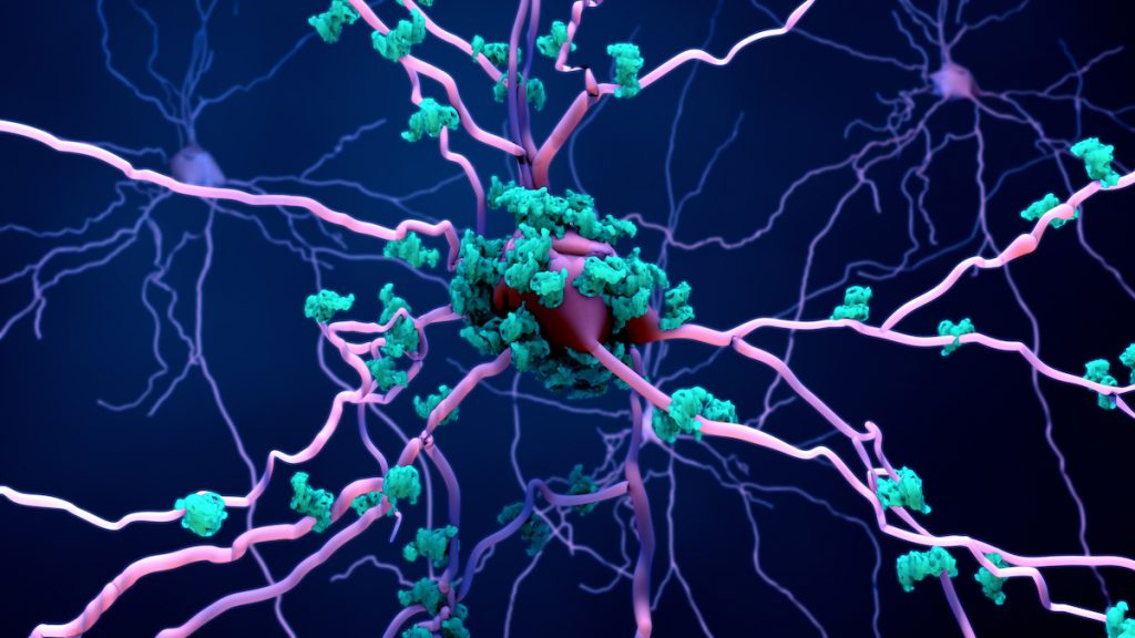
The research identified a specific neuronal population in FCD patients characterized by the expression of NEFM+ neurofilament (a neuronal protein), which includes the so-called dysmorphic neurons. These are abnormal cells found in the cortex of these patients that are responsible for the altered synapses that cause epileptic seizures.
As for microglia (immune system cells located in the brain), two subpopulations were discovered, called CD74+ and CD83+. These may be associated with immune activation and neuroinflammation.
“Using an advanced genomic technique, we obtained a cellular and therefore extremely detailed view of this brain malformation,” said computational biologist Diogo Veiga, corresponding author of the article.
“We identified profound cellular changes in the cortex of these patients, including the loss of neurons in the upper layers, as well as immature astrocytes and populations of microglia expanded in the lesions and associated with inflammation. This cell atlas is of great importance for understanding the mechanisms and seeking specific therapies that can target the identified cells.”
In work that began in 2021, the group generated a dataset of 61,525 single cells from 11 clinical samples of focal cortical dysplasia lesions obtained from patients undergoing surgery and from controls.
Single-cell sequencing is an advanced molecular biology technique that allows genetic material (DNA or RNA) to be analysed individually, providing a detailed view of cellular heterogeneity and revealing much more specific characteristics of the lesions studied.
One of the challenges was analysing the amount of data.
“We spent a long time developing the computational workflow and testing different approaches to be able to identify these subpopulations associated with the disease,” said Veiga, who highlights the contribution of his PhD thesis student Isabella Cotta Galvão, first author of the article.