Stroke care has always been a race against time, and MRI – despite its precision – often slows that race down when every minute matters. Radiologists know this well: a patient arrives, several sequences run one after another, and then a stack of images waits for interpretation.
AI in functional MRI is no longer a future promise; it is becoming a practical tool for identifying ischemic changes, predicting tissue viability, and supporting emergency diagnoses with speed and consistency.
Why Speed Matters in MRI-Based Stroke Detection
A stroke develops rapidly. Within minutes of a vessel occlusion, neuronal tissue begins to fail. MRI can detect early microstructural and metabolic disruptions far more sensitively than CT; however, its diagnostic precision does not eliminate a crucial bottleneck: time.
Key delays often occur due to:
- Complex sequence interpretation (DWI, ADC maps, perfusion imaging, fMRI-based hemodynamic signals)
- Limited access to neuroradiology experts during night shifts or in smaller hospitals
- Large case volumes leading to reading queues
Ambiguous early-stage patterns that require nuanced assessment
AI in functional MRI addresses each of these points by automating detection, highlighting abnormalities, and quantifying patterns that typically require years of clinical experience.
Problem: Diagnostic Challenge Under Time Pressure
Standard MRI diagnostics require time – not only for the scanning procedure itself but also for subsequent interpretation by a radiologist. In cases of acute traumatic brain injury (TBI), the situation can be complex: a combination of brain contusion, edema, possible hemorrhage, and ischemia (impaired blood flow). Even the most experienced human eye can miss subtle signs – small but critically important indicators of early ischemia amidst traumatic changes.
The challenge becomes even greater when a patient arrives at a small hospital without round-the-clock coverage by a specialized neuroradiologist. The need to transfer images to another center or wait for a specialist can consume precious minutes of the “therapeutic window.”
Solution: Algorithm as an Expert Consultant. How AI Implements Rapid MRI Analysis
The shift toward automated MRI interpretation has accelerated due to advances in deep neural architectures specifically adapted for brain imaging. Modern systems, trained on large brain MRI datasets, combine convolutional neural networks, 3D image transformers, and spatiotemporal modeling frameworks capable of processing the high dimensionality of functional MRI data. Instead of evaluating images slice by slice, these models examine entire volumes, track perfusion dynamics over time, and interpret subtle BOLD (blood-oxygen-level-dependent) fluctuations that often escape rapid manual review.
Core Capabilities
Lesion Segmentation
Algorithms outline ischemic regions automatically, generating volumetric masks based on DWI abnormalities, ADC reductions, and functional disruptions. Segmentation informs both initial diagnosis and longitudinal monitoring.
Perfusion Deficit Detection
Perfusion MRI generates multiple hemodynamic maps—MTT, CBF, CBV, TTP—each sensitive to a different element of blood flow. AI systems evaluate these sequences as a unified dataset, detecting areas with delayed or insufficient perfusion. Functional MRI inputs add context by showing how affected regions respond at the metabolic level.
Mismatch Quantification
Mismatch represents the difference between irreversibly damaged core tissue and potentially viable penumbra. Automated quantification accelerates decisions on thrombolytic therapy and endovascular intervention. Models provide volumetric calculations that traditionally require manual tracing and cross-sequence comparison.
Diffusion Abnormality Scoring
Diffusion-restricted areas often appear early in ischemia, but subtle changes can be difficult to characterize quickly. Automated scoring systems assign numerical severity values based on texture patterns, shape, and ADC depression, enabling standardized assessment across patients and institutions.
Predictive Outcome Modeling
Outcome prediction models use lesion topography, size, perfusion deficits, and disrupted functional connectivity patterns to estimate neurological recovery potential. These estimates assist in planning treatment intensity and setting expectations for rehabilitation.
Workflow Prioritization
Triage modules automatically push high-risk cases to the top of reading queues. Instead of waiting for manual sorting, scans with suspected vessel occlusion receive immediate attention, reducing time-to-treatment.
How the Models Interpret Data
AI systems for stroke diagnostics typically follow a multi-stage pipeline:
- Preprocessing – Motion correction, normalization, skull stripping, and noise reduction across both structural and functional sequences.
- Feature Extraction – Identification of diffusion signatures, perfusion time-intensity curves, and fMRI BOLD variations.
- Spatial-Temporal Fusion – Integration of 3D anatomical structures with temporal perfusion data.
- Segmentation and Scoring – Isolation of key abnormalities and quantification of lesion metrics.
- Decision Layer – Estimation of stroke subtype, extent, and recommended urgency level.
This pipeline transforms raw MRI volumes—often hundreds of slices across multiple time points—into structured, clinically actionable information.
Functional Contributions of Key MRI Sequences in AI-Assisted Stroke Analysis
| MRI Sequence | Physiological Indicator | AI-Extracted Insights | Clinical Relevance |
| DWI | Water diffusion restriction | Identification of acute cytotoxic edema | Early ischemic core detection |
| ADC Map | Quantitative diffusion values | Severity scoring and artifact suppression | Confirmation of true diffusion restriction |
| Perfusion MRI (MTT, CBF, CBV) | Blood flow and transit dynamics | Perfusion deficit segmentation and mismatch estimation | Penumbra identification and revascularization suitability |
| T2-FLAIR | Fluid and tissue contrast | Chronological stage differentiation | Helps distinguish acute from subacute lesions |
| Functional MRI (BOLD) | Hemodynamic and metabolic response | Detection of disrupted connectivity and stress patterns | Early functional impairment before structural change |
Rapid MRI Analysis for Acute TBI: A Brief Example
Patient scenario:
A patient arrives, several sequences run one after another, and then a stack of images waits for interpretation
How Rapid Analysis Works
Scanning (5–7 minutes). A standard, shortened MRI protocol is performed.
Image Upload. The system receives DICOM files immediately from the scanner.
Automatic Processing (30–60 seconds)
- Noise reduction
- Image alignment
- 3D model reconstruction
Pathology Detection
The algorithm highlights suspicious areas:
- Hematomas
- Microbleeds
- Ischemic regions
- Midline shift
Final Report (10–15 seconds)
The clinician receives a damage map, hematoma volume, risk of herniation, and deviations from normal structures.
Total time: 2–3 minutes after scanning.
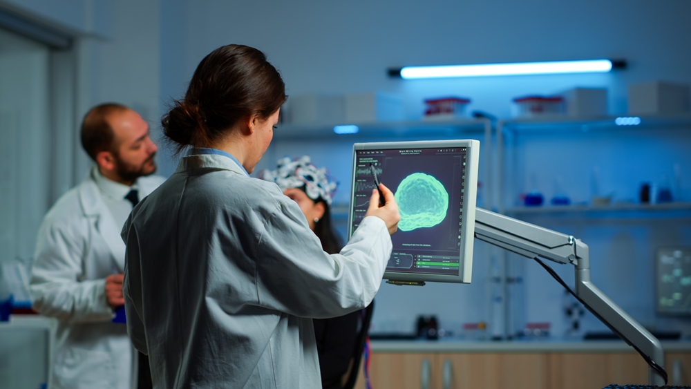
Researcher looking at monitor analysing brain scan while coworker discussing with patient in background about side effects, mind functions, nervous system, tomography scan working in laboratory
Comparison of Traditional MRI Analysis vs AI-Assisted Rapid Analysis
| Parameter | Without AI (Traditional Mode) | With AI (Rapid Analysis) |
| Image Review | Clinician manually scrolls through dozens of slices | Algorithm analyzes the entire volume within seconds |
| Microdamage Detection | Manually searches for microbleeds and small lesions | Automatically highlights suspicious areas, including subtle and hard-to-see ones |
| Segmentation & Volume | Performed manually, compared with previous scans | Builds a 3D damage map and calculates volumes |
| Report & Interpretation | Clinician prepares conclusions independently | Ready-made summary for clinician review |
| Error Risk | Higher, especially under a heavy workload | Lower, fatigue-related errors are reduced |
| Decision Time | 8–15 minutes or more | 1–3 minutes, decisions made 3–5 times faster |
Effectiveness of Early Stroke Diagnosis Using AI
AI-based systems for early stroke diagnosis have been used in medicine for several years. Among them are services such as “CT Brain” (Russia), Rapid, Viz LVO (USA), Brainomix (UK), and Avicenna CINA (France). Based on research, these systems demonstrate high accuracy rates — over 80%.
These AI systems assist clinicians by rapidly analyzing brain imaging, detecting signs of ischemic or hemorrhagic stroke, and prioritizing urgent cases. Their use has been shown to significantly reduce the time to diagnosis and treatment, which is critical in improving patient outcomes. Moreover, integration of AI into stroke care pathways supports decision-making, helps optimize resource allocation, and can potentially decrease long-term disability associated with delayed intervention. Ongoing studies continue to evaluate their effectiveness across different populations and healthcare settings, aiming to further refine algorithms and expand accessibility.
Artificial intelligence is an effective tool for early stroke diagnosis and severity assessment. It shortens the time to medical intervention and helps physicians make correct clinical decisions. AI-based stroke diagnostic systems have been developed and implemented in most developed countries. Numerous studies confirm the benefits of AI in clinical practice .
Conclusion
AI in functional MRI is reshaping the speed and precision of stroke diagnostics. Automated analysis delivers rapid lesion detection, objective quantification, and consistent insights across hospitals of all sizes. From triage to treatment planning, AI reduces delays that once cost patients valuable time. As imaging datasets expand and algorithms grow more sophisticated, rapid MRI interpretation is evolving into a standard component of acute care.
If adopted widely, these systems have the potential to reduce disability, standardize care across regions, and support clinicians in making faster, more informed decisions at the exact moment they matter most.