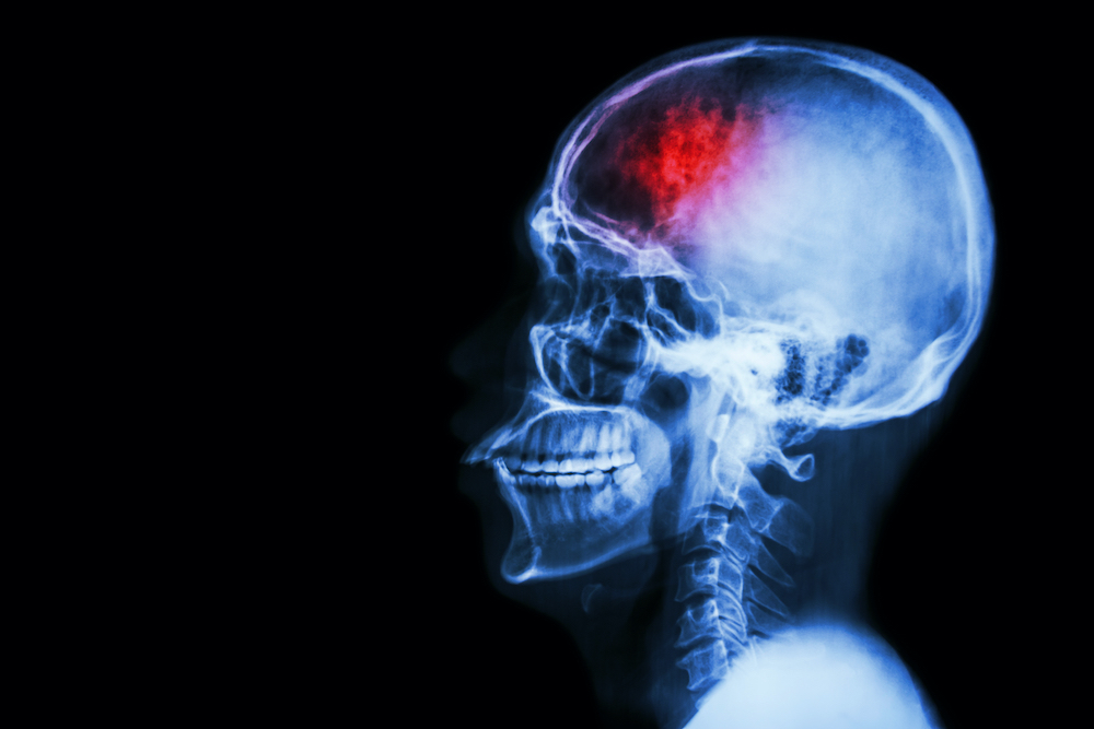
According to a new study, researchers have gained new insight on how the brain can repair itself post-stroke.
Functional decline after an ischaemic stroke is a common experience for patients, especially due to the brain’s resistance to regenerate after damage. However, there is still potential for recovery as surviving neurons can activate repair mechanisms to limit and even reverse the damage caused by the stroke. The question this study looked to answer is how is it triggered?
Researchers from Tokyo Medical and Dental University (TMDU) provided new insight to this question by identifying a new mechanism. They discovered that neurons surrounding the area of cell death secrete lipids that can trigger brain-autonomous neural repair after ischaemic brain injury.
An ischaemic stroke occurs when the blood supply to the brain is blocked and results in the death of brain cells. This condition is life-threatening, and patients will likely develop functional disabilities. Although the adult brain can self-repair, the underlying mechanisms need further clarification.
Inflammation of the brain contributes to the effects of ischaemic stroke.
Senior author, Takashi Shichita, says: “There is evidence that more lipids are produced after tissue injuries and contribute to regulating inflammation.
“We investigated the changes in lipid metabolite production in mice after ischaemic stroke. Interestingly, the levels of a specific fatty acid called dihomo-γ-linolenic acid (DGLA) and its derivatives increased after the stroke.”
The researchers further discovered that a protein called PLA2GE2 (Phospholipase A2 Group IIE, an enzyme) mediates DGLA increase. By manipulating the expression of PLA2GE2, they also displayed its impact on functional recovery. Deficiency of PLA2GE2 caused more inflammation, lower expression of factors stimulating neuronal repair, and more tissue loss. The team carried on with identifying the targets of PLA2GE2/DGLA.

Lead author of the study, Akari Nakamura, says: “When we look at genes expressed in mice lacking PLA2GE2, we found low levels of a protein called peptidyl arginine deiminase 4 (PADI4).
“PADI4 regulates transcription and inflammation. Remarkably, expressing PADI4 in mice limited the extent of tissue damage and inflammation after ischaemic stroke!” Additionally, the study shows that PADI4 promotes the transcription of genes involved in brain repair. It also identifies the whole signaling pathway involved in this process.”
Most data was obtained in a mouse model of ischaemic stroke. Yet, the recovery pathway likely exists in humans as the researchers found that neurons surrounding the stroke site express PLA2G2E and PADI4 in humans. Moreover, another recent study reported that the lower serum DGLA level was correlated with the severe ischaemic stroke and cognitive disorders in humans.
This study describes a new mechanism that triggers brain repair after an ischaemic stroke, which might lead to the development of compounds promoting PADI4’s effects, that stimulate the recovery of patients. It could also change our current understanding and approach toward Eicosapentaenoic acid (EPA) or Docosahexaenoic acid (DHA), as the only beneficial lipids for preventing atherosclerosis and vascular diseases.