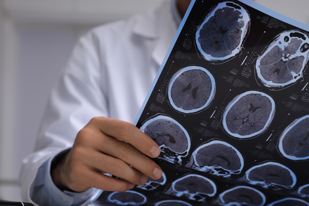
AI has revealed previously undetectable brain lesions in people with MS, in research that could change how the condition is monitored.
Multiple sclerosis, commonly known as MS, is a debilitating condition in which the immune system mistakenly attacks the nervous system. It affects around 150,000 people in the UK.
It can cause fatigue, vision problems, muscle spasms, memory difficulties and balance issues. Lesions and scarring in the brain and spinal cord are also hallmarks of the condition.

Scientists have long known that damage to grey matter, which supports information processing, movement, memory and emotion, is a major driver of disease progression.
However, the MRI scans used to monitor people with MS have traditionally detected lesions in the brain’s white matter rather than its grey matter.
This has left researchers with few ways to track hidden grey matter damage. Many MS drugs developed in recent years have therefore focused on treating white matter lesions.
Scientists at the University at Buffalo have now developed a way to reveal grey matter lesions, which they say could have implications for MS treatment and research.
Robert Zivadinov, senior author and professor of neurology at the University at Buffalo, said: “Detecting previously invisible cortical lesions on conventional legacy MRI scans has major implications for MS research and clinical care.
“The ability to see for the first time these previously hidden indicators of MS disease progression, including cognitive impairment and disability, is an important advance.”
The researchers used artificial intelligence to compare several different types of MRI image from the same brain simultaneously.
They tested the technique on scans from more than 700 people with MS and uncovered more than 11,000 previously undetectable grey matter lesions.
Although each scan showed little or no sign of grey matter damage on its own, the AI identified small differences between the images that could not be seen by the human eye.
The researchers said the technology could also identify areas where brain tissue was not behaving like healthy tissue.
Michael G. Dwyer, first author and associate professor of neurology and biomedical informatics in the Jacobs School, said: “We have all been very frustrated, knowing that these cortical lesions were there but not being able to see them.
“There’s a lot of ongoing damage that continues to happen in MS that you won’t see with conventional MRI, but that histopathologists have been clearly demonstrating for decades on postmortem tissue.
“What this collaboration has been able to accomplish is a real success story for applying AI in the medical arena.
“We now have access to these incredibly useful data on MRI scans that were there but you couldn’t see them without using AI to pull them out. The computational methods are finally at the point where we can do this.”
Histopathologists are specialists who examine tissue under a microscope to identify changes caused by disease or injury.
MS lesions are thought to cause symptoms including weakness, numbness, vision problems and difficulty with balance.
They disrupt the brain’s communication network by slowing or blocking nerve signals.
Zivadinov added: “This work, which has revealed that there is so much invisible pathology in the brain, will have tremendous impact for reviewing data from past clinical trials and also for those going forward.”
The number of people living with MS in Britain has risen by around 20,000 since 2019.
Symptoms usually first appear between the ages of 20 and 40. The condition is thought to develop when the immune system attacks myelin, the protective coating around nerve fibres, causing inflammation and damage to the central nervous system.
MS is not usually fatal, but advanced disease can weaken the muscles used for breathing and swallowing and increase the risk of serious infections.
There is no cure, but treatments can slow the progression of the disease.