From robotics and Virtual Reality (VR) to precision rehabilitation, NR Times speaks to Professor Stephanie Clarke of The World Federation for Neurorehabilitation (WFNR) about the latest research developments and challenges in the world of cognitive rehabilitation.
WFNR is a multidisciplinary organisation based in the UK that drives awareness of neurorehabilitation across the globe, providing training and education, and encouraging research and collaboration.
Professor Clarke, of the Neuropsychology and Neurorehabilitation Department at CHUV, Lausanne, Switzerland, is head of the WFNR’s Cognitive Rehabilitation Special Interest Group which works to promote an integrated approach to cognitive disorders.
The group focuses on cognitive interventions from the acute to chronic stages, indications for behavioural and pharmacological treatments, and evidence-based approach to treatment of cognitive disorders.
Professor Clarke spoke to NR Times about the latest advancements in neuro rehabilitation, including robotics, virtual reality, neuroimaging and precision rehabilitation.
Can you give me an overview of current research advances?
There are new advances in behaviour interventions as well as in neuroimaging, understanding what’s happening in the brain when we do rehab and how we can use it for patients.
Brain Stimulation is also something which is in the pipeline as well as robotic approaches. Some of them are in clinical practice, but some of them are really in between. Of course, there is always the hope that we will be able to use cellular and molecular approaches, but they are more in basic research and some in translational research for the moment.
In what ways are robotics advancing the field of cognitive rehabilitation?
There are now robotics which help patients to train. There are walking robots which help patients to do a lot of walking, train their gait or retrain arm movements – they are used for rehabilitation instead of a physiotherapist accompanying each movement.
Then you have robotics which help people to become independent. For example, an exoskeleton which supports somebody who has lost muscular function to a certain degree and helps take the weight off the leg so that the patient can walk.
There are also very sophisticated robots where you make an interface with the brain which can help paralytic patients walk again. They use brain signals to drive implants in the spinal cord so that the patients can start walking again. This is a fascinating field for the moment, but cannot be applied to every patient.
For these technologies, patients are selected who have the best chance of succeeding.
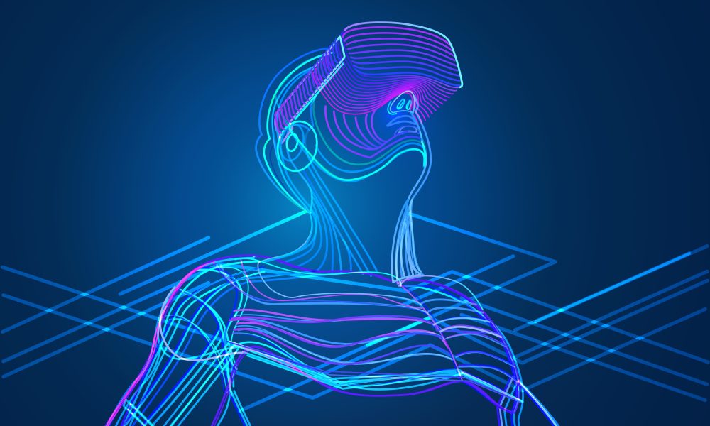
How is VR advancing cognitive neurorehabilitation?
Virtual Reality is able to train a lot more functions because in the real world, it’s less motivating.
With virtual reality, you can make it more attractive. You can also change visual motor perception in virtual reality in a very conformed way, improving certain aspects like tension.
There is a condition called neglect, where patients do not pay attention to things on the left side. If you shift the visual world for a while you can reorganise the brain so that they are able to pay attention to the things on the left.
Previously this was done with presence glasses which deviate. Now you can start doing this in virtual reality, which is much more adaptable to clinical practice, for example, when the patients are not at the hospital, when they are far away and cannot go to for the treatments every day.
Are there any challenges with the application of these technologies?
There are some issues. In medicine, you have clinical trials which are randomised and controlled to show that this approach is better than the control.
However, in neuro rehabilitation, and especially for cognitive functions, there are critical dividends for some, but for some, the evidence is not convincing. And we were wondering why it
There are a series of examples where the application of a rehabilitation approach of training or intervention may not be best for patients who have slightly similar problems. For example, with the neglect problem of patients not paying attention to the left side, there are different approaches, but when they are run in randomised control trials, they are not very convincing.
Our group has started to look at why that could be, so we looked into brain mechanisms. We found that when you expose the patients to this visual motor deviation, you make them adapt, you change brain organisation so that they can use a compensatory system in the brain for further attention.
Now, there are patients in whom this compensatory system is preserved and there are others where it’s damaged as well. It’s logical that then this therapeutic approach cannot help them, however, there are others where there are patients who will respond to a treatment and those who don’t.
Usually it depends on what the issue is, and what it destroys. This is a very important understanding because the danger was that, because of these negative results in clinical trials, a whole series of treatments would be thrown away and patients will not be able to benefit from that.
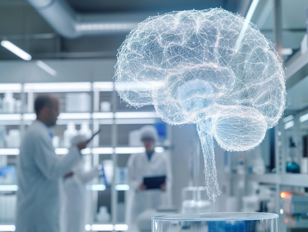
Are there any notable advances in neuroimaging?
There are basically two types of studies in patients – cognitive functions for instance, or motor functions. We know how it works in normal subjects or what is activated if we do certain movements with our hand or when we speak or read and so on, and this is reorganised in the patients.
One is understanding how this normal functioning breaks down these specific regions and how the deficits reflect in this reorganisation. The next question is, what is the best prognosis if a patient has, for instance, a left hemispheric lesion. Some patients will recover and some don’t.
Now, there are studies which help us to understand recovery and brain plasticity. In individual patients there is a limited use of functional imaging.
There are a few conditions in which we know that the recovery will not take place because the critical structures which could help with the recovery have been destroyed by the lesion. Instead of engaging in weeks of rehabilitation, there is a possibility to do good brain imaging to know where the lesion is to see if some aspects of cognitive function have been disturbed.
For instance, some patients have a deficit in understanding language. Many recover but some don’t, because there are very critical lesions. In these patients, it is worthwhile to start other types of communication so that they can get used to it, use it with their families, and so on. Work on better compensation instead of trying to rehabilitate something which will not come.
Other aspects that help us with brain imaging is what we call personalised medicine, or precision medicine. With the imaging data and very precise history of the patient, we can design a rehabilitation path for this specific patient. Brain imaging contributes a lot to that.
I think precision rehabilitation is something which we need to consider, and very likely it will be pushed further with cellular, perhaps even molecular indicators.