‘Irreversible’ spinal cord damage may be reversed, research suggests
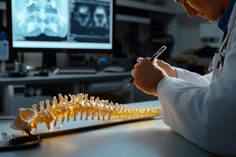
Spinal cord damage once thought irreversible may be reversible, after researchers grew miniature brain and spinal circuits in the lab.
The early-stage research suggests nerve fibres connecting the brain and spinal cord may be able to regrow after damage if key biological signals are targeted.
The findings could help scientists understand why paralysis and other disabilities after central nervous system injury are often permanent.

Senior author Dr András Lakatos, who led the project at the University of Cambridge department of clinical neurosciences, said: “When the brain and spinal cord are damaged, the nerve fibres that carry movement signals from the brain to the spinal cord rarely grow back.
“That’s why paralysis is usually permanent. But we didn’t know exactly when the ability of axons to regenerate becomes limited.
“Our model provides a good indication that this block happens during development, and it can still be reversed after this point.”
Researchers at the University of Cambridge developed miniature lab-grown circuits that mimic how the brain and spinal cord connect to control movement.
The circuits were made using organoids, which are small 3D structures grown from stem cells to resemble parts of human tissue.
As people develop, nerve cells, known as neurons, form connections that allow messages to pass between the brain, spinal cord and muscles.
A key part of each neuron is the axon, a long nerve fibre that carries signals to other cells.
At some point during development, the central nervous system largely loses its ability to regrow axons after injury.
This means damage to the brain or spinal cord can become permanent, causing disabilities such as loss of hand movement or the ability to walk.
This can happen after traumatic spinal cord injury and may also be a feature of neurological conditions including motor neurone disease and multiple sclerosis.
Motor neurone disease affects the nerve cells that control movement, while multiple sclerosis is a condition in which the immune system damages the brain and spinal cord.
In 2021, Dr András Lakatos and colleagues developed “mini brains” using stem cells derived from human patients.
The team has now built a miniature connected brain and spinal cord system in the lab using organoids.
In the human body, the brain and spinal cord are separate but connected by axons, so the researchers kept the brain and spinal cord organoids apart.
They found that nerve fibres from the brain tissue grew across the gap to connect with the spinal cord tissue, creating a working circuit that could trigger tiny muscle clusters to contract.
By growing the human system in a dish for more than a year, the team found that axons could regrow after damage until around day 150.
This stage corresponds to the middle trimester of pregnancy.
After that point, axon growth was greatly impaired.
George Gibbons, from the department of clinical neurosciences at the University of Cambridge and first author of the study, said: “Neurons taken from less mature organoids regrew long fibres after injury, but those from more mature organoids showed a sharp drop in their ability to regrow.
“In other words, poor regeneration is built into human neurons as they mature in the central nervous system.”
The researchers analysed gene expression, which shows how active genes are within cells.
They identified a network of genes that appears to act as a switch, restricting axon growth as neurons mature and form synapses.
Synapses are the connections that allow nerve cells to communicate with each other.
The team found that blocking key regulators of this network switched axon growth back on.
They then searched a database of drug compounds for substances that act on the genes involved and identified lynestrenol, a hormone drug licensed for some menstrual disorders and as a contraceptive.
When tested on damaged neurons, the drug significantly increased axon regrowth.
The researchers said lynestrenol itself may not be the answer to spinal cord repair, but could point towards ways to directly target human neurons.
Scar tissue and inflammation may also limit axon repair after injury, but the team said understanding neuron-specific causes is important.
They said this is supported by evidence that axons from less mature neurons can grow through difficult injury-site environments.
Organoid models are increasingly used to study human biology and disease.
While animal models such as mice and rats can help researchers understand some biological processes, organoids grown from human stem cells can more closely mimic human tissue.
Dr Lakatos said: “Much of what we know about nerve regeneration comes from rodents, whose neurons behave differently from human neurons.
“Our sophisticated organoid models help bridge the knowledge gap from animal models to what we see in patients. They are also an important contribution to efforts to reduce the use of animals in research.”
Organoids, often described as “mini organs”, are being used increasingly to model human biology and disease.
At the University of Cambridge, researchers use them to study areas including damaged livers, Crohn’s disease in children and the early stages of pregnancy.