Researchers to create AI models for early prediction of Alzheimer’s
Researchers in the US have received a National Institutes of [...]
Researchers in the US have received a National Institutes of [...]
In an early new year boost for Calvert Reconnections, case [...]
Researchers have discovered that a protein called phosphorylated α-synuclein, which [...]
The value of the global market for rehab robotics is [...]
The assistive planar robot includes a closed-loop feedback system to monitor the muscle and brain activity of the user in order to trigger the execution of reach and grab in an adaptive way.
In a new paper, researchers at the University of Rhode Island Motor Control and Rehabilitation lab write: “Numerous rehabilitation approaches such as muscular electrical stimulations, brain-computer interfaces, and transcranial magnetic stimulation have been investigated to assist the affected individuals.
These lab-grown organoids open up a brand-new way of studying how the brain develops. They also offer a valuable means to study the development and treatment of diseases related to brain development, including brain tumours.
Scientists use different ways to model the biology of healthy tissue and disease in the lab. These include cell lines, laboratory animals and, since a few years, 3D mini-organs.
These so-called organoids have characteristics and a level of complexity that allows scientists to closely model the functions of an organ in the lab.
Organoids can be formed directly from cells of a tissue. Scientists can also ‘guide’ stem cells – found in embryos or in some adult tissues – to develop into the organ they aim to study.
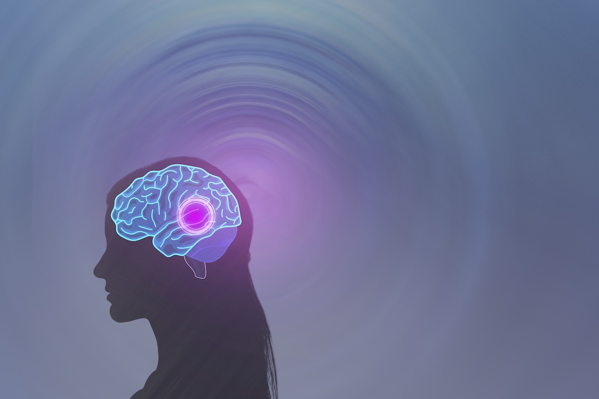
In the development of Parkinson’s, the changes that will lead to neurodegeneration take place in the brain long before patients show any symptoms.
But without a test that can detect these changes, it’s difficult to intervene early to more effectively slow disease progression.
Doctors generally must rely on neurological examination and patients’ medical histories when diagnosing Parkinson’s.
Once clinical symptoms appear, however, the disease has already wreaked irreversible damage in the brain.
Researchers at Weill Cornell Medicine have catalogued the cellular response to stroke in a preclinical model, identifying the immune cells involved and the roles they may play in the days and weeks following a stroke.
During a stroke, loss of oxygen leads to brain damage and cell death. It also triggers a powerful inflammatory response in which the brain’s resident immune cells, along with cells recruited from the blood, infiltrate the injured tissue.
Boston University's neuro-rehab centre has published an in-depth account of [...]
Other Aspect Health Media Titles
![]()
![]()
![]()
Copyright © 2025 Aspect Health Media Ltd